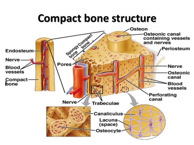
Compact Bone Diagram ~ Compact Bone Diagram Microscope Compact Bone Diagram Koibana Info Anatomy And Physiology Human Anatomy And Physiology Anatomy Bones Sclerostin Inhibits Bone Formation Mostly By Antagonizing Lrp5 6 Thus Inhibiting Wnt Signaling. There are two types of bone tissue: The diagram above shows a longitudinal view of an osteon. Compact bone diagram osteon compact bone ap pinterest anatomy human anatomy and. The two layers of compact bone and the interior spongy bone work together to protect the internal organs. Diagram of a typical long bone showing both cortical (compact) and cancellous (spongy) bone.
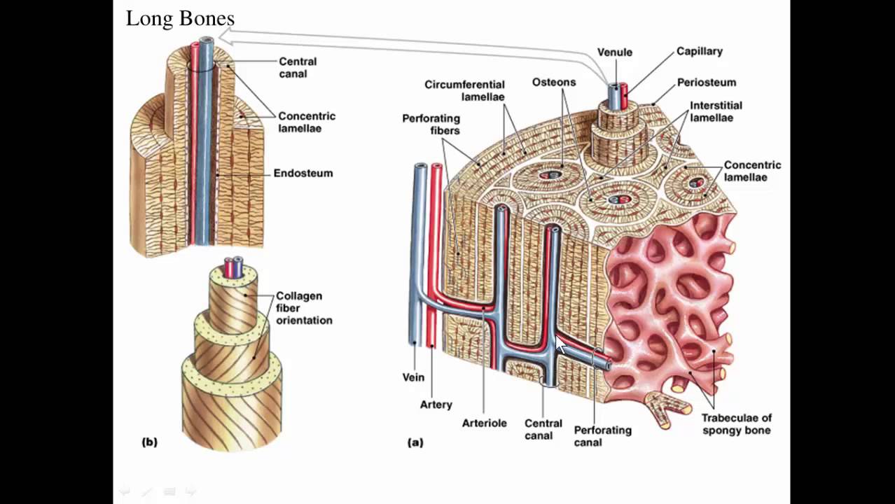
Anatomy of shoulder 12 photos of the anatomy of shoulder anatomy of nerves in shoulder, anatomy of posterior shoulder dislocation, anatomy of right shoulder, anatomy of shoulder labrum tear, anatomy of the shoulder games, human anatomy, anatomy of nerves in shoulder, anatomy of posterior shoulder dislocation, anatomy of. Compact bone is the denser stronger of the two types of bone tissue. (b) in this micrograph of the osteon, you can clearly see the concentric lamellae and central canals. The two layers of compact bone and the interior spongy bone work together to protect the internal organs. About press copyright contact us creators advertise developers terms privacy policy & safety how youtube works test new features press copyright contact us creators.
Histo Bone from image.slidesharecdn.com As the bony trabecullae increase in width and length by addition of new lamellae, all the mesenchyme is replaced by cancellous bone that is transformed into compact bone later. Compact bone is the denser stronger of the two types of bone tissue. Compact bone is the strongest form of bone tissue containing few spaces. These bones are tough and hard with negligible gaps inside them. The compact bone is a dense bone found in the diaphysis. The cells of compact bone, which is also called cortical bone, appear to be tightly packed into a solid mass. Cortical bone is compact bone while cancellous bone is trabecular and spongy bone. Add to favorites 0 favs.
Anatomy of shoulder 12 photos of the anatomy of shoulder anatomy of nerves in shoulder, anatomy of posterior shoulder dislocation, anatomy of right shoulder, anatomy of shoulder labrum tear, anatomy of the shoulder games, human anatomy, anatomy of nerves in shoulder, anatomy of posterior shoulder dislocation, anatomy of.
The shafts found in long bones are also compact bones. Compact bone is formed from a number of osteons, which are circular units of bone material and blood vessels. (b) in this micrograph of the osteon, you can clearly see the concentric lamellae and central canals. Compact and spongy tissues in a flat bone. It makes up the outer cortex of all bones and is in immediate contact with the periosteum. Compact bone is the denser, stronger of the two types of osseous tissue (figure 6.3.6). The cells of compact bone, which is also called cortical bone, appear to be tightly packed into a solid mass. Its repeated pattern is arranged in concentric layers of solid bone tissue. Contrary to compact bone, spongy bone or. Cortical bone is compact bone while cancellous bone is trabecular and spongy bone. Related posts of compact bone diagram labeled anatomy of shoulder. The remainder is cancellous bone, which has a spongelike appearance with numerous large spaces and is found in the. Compact bone, also called cortical bone, is the hard, stiff, smooth, thin, white bone tissue that surrounds all bones in the human body.
The two layers of compact bone and the interior spongy bone work together to protect the internal organs. Compact bone, also called cortical bone, dense bone in which the bony matrix is solidly filled with organic ground substance and inorganic salts, leaving only tiny spaces (lacunae) that contain the osteocytes, or bone cells.compact bone makes up 80 percent of the human skeleton; The two main structural components typically include spongy bone on the interior, with an outer layer of compact bone. Nov diagram for.net is a fully managed, extensible and powerful diagramming framework, which can help you create feature rich diagramming solutions in winforms, wpf, silverlight, xamarin.mac. Anatomy of shoulder 12 photos of the anatomy of shoulder anatomy of nerves in shoulder, anatomy of posterior shoulder dislocation, anatomy of right shoulder, anatomy of shoulder labrum tear, anatomy of the shoulder games, human anatomy, anatomy of nerves in shoulder, anatomy of posterior shoulder dislocation, anatomy of.
Diverzija Pravedan Europa Compact Bone Thehoneyscript Com from i.ytimg.com In long bones, as you move from the outer cortical compact bone to the inner medullary cavity, the bone transitions to spongy bone. It is also called osseous tissue or cortical bone and it provides structure and support for an organism as part of its skeleton, in addition to being a location for the storage of minerals like calcium.about 80% of the weight of the human skeleton comes from. (b) in this micrograph of the osteon, you can clearly see the concentric lamellae and central canals. These bones are tough and hard with negligible gaps inside them. Thin layer of reticular ct lining internal marrow cavity. As the bony trabecullae increase in width and length by addition of new lamellae, all the mesenchyme is replaced by cancellous bone that is transformed into compact bone later. Compact bone, as opposed to spongy bone, is made of cylindrical units, called osteons, that are tightly formed together. Between the rings of matrix the bone cells osteocytes are located in spaces called lacunae.
Compact and spongy tissues in a flat bone.
Haversian canals (sometimes canals of havers) are a series of microscopic tubes in the outermost region of bone called cortical bone. Compact bone is the strongest form of bone tissue containing few spaces. There are small canals that run through the bone, which allow blood vessels to penetrate it. The shafts found in long bones are also compact bones. Although the calls are close together, this type of bone is not completely solid. Thin layer of reticular ct lining internal marrow cavity. The compact bone can be seen as the layer just underneath the periosteum, color both ends. Some, mostly older, compact bone is remodelled to form these haversian systems (or osteons). Provides protection and support while resisting stress from weight and movement. Its repeated pattern is arranged in concentric layers of solid bone tissue. Under periosteum of all bones is the bulk of the diaphysis of long bones. Compact bone tissue diagram quizlet. Compact bone diagram osteon compact bone ap pinterest anatomy human anatomy and.
The remainder of the bone is formed by cancellous or spongy bone. It makes up the outer cortex of all bones and is in immediate contact with the periosteum. It is also called osseous tissue or cortical bone and it provides structure and support for an organism as part of its skeleton, in addition to being a location for the storage of minerals like calcium.about 80% of the weight of the human skeleton comes from. There are pores and spaces even in compact bone. Compact bone diagram osteon compact bone ap pinterest anatomy human anatomy and.
Osteoporosis Anatomy from www.spineuniverse.com About press copyright contact us creators advertise developers terms privacy policy & safety how youtube works test new features press copyright contact us creators. Microscopic structures of compact bone wedge of bone duration. There are two types of bone tissue: Compact bone is the denser, stronger of the two types of osseous tissue (figure 6.3.6). Similarities between compact bone and spongy bone @. Some, mostly older, compact bone is remodelled to form these haversian systems (or osteons). Add to favorites 0 favs. They are soft and light bones make up of loosely packed trabeculae.
Nov diagram for.net is a fully managed, extensible and powerful diagramming framework, which can help you create feature rich diagramming solutions in winforms, wpf, silverlight, xamarin.mac.
Compact bone, also called cortical bone, is the hard, stiff, smooth, thin, white bone tissue that surrounds all bones in the human body. They allow blood vessels and nerves to travel through them to supply the osteocytes Compact bone, also called cortical bone, dense bone in which the bony matrix is solidly filled with organic ground substance and inorganic salts, leaving only tiny spaces (lacunae) that contain the osteocytes, or bone cells.compact bone makes up 80 percent of the human skeleton; Diagram of a typical long bone showing both cortical (compact) and cancellous (spongy) bone. Human bone generally comprises osseous tissue, an outer coating called a periosteum, and bone marrow. Contrary to compact bone, spongy bone or. Related posts of compact bone diagram labeled anatomy of shoulder. Compact bone is the denser stronger of the two types of bone tissue. Although the calls are close together, this type of bone is not completely solid. The compact bone can be seen as the layer just underneath the periosteum, color both ends. These bones are tough and hard with negligible gaps inside them. Add to favorites 0 favs. Nov diagram for.net is a fully managed, extensible and powerful diagramming framework, which can help you create feature rich diagramming solutions in winforms, wpf, silverlight, xamarin.mac.